Request for Funding for Scientific Events text 2023-2024
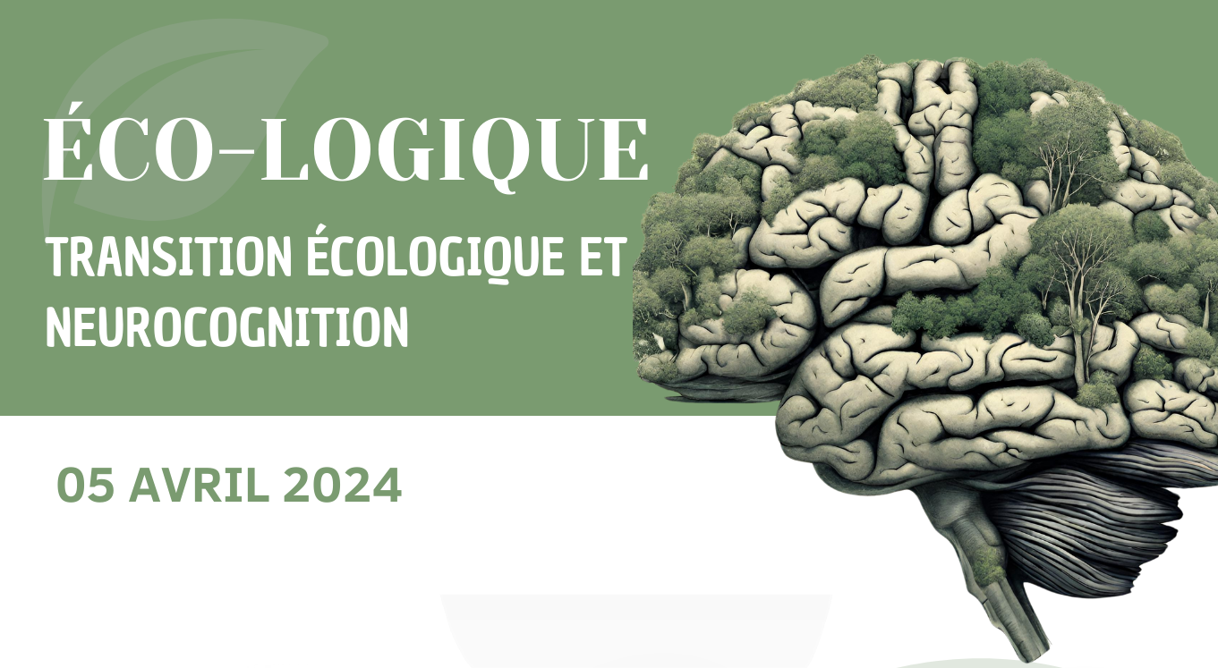
The CerCoG program is organizing its interdisciplinary scientific seminar entitled "Eco-logic: Ecological Transition and Neurocognition" on April 5th from 8:30 am to 1:00 pm.
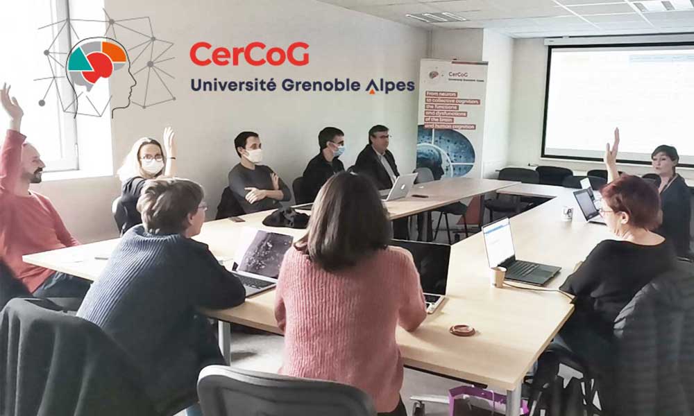
CerCoG's scientific events
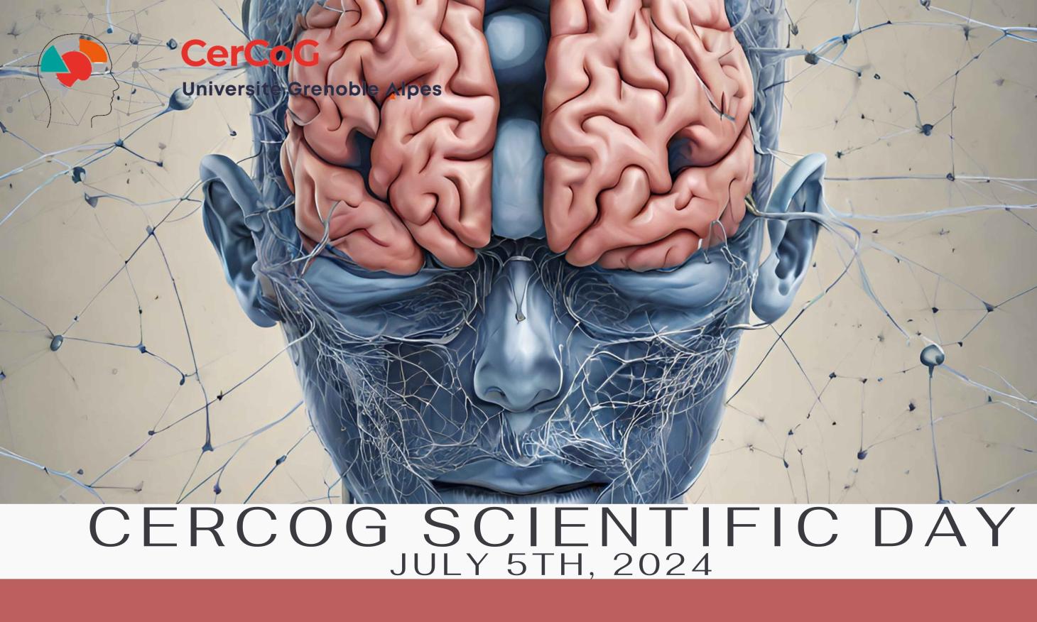
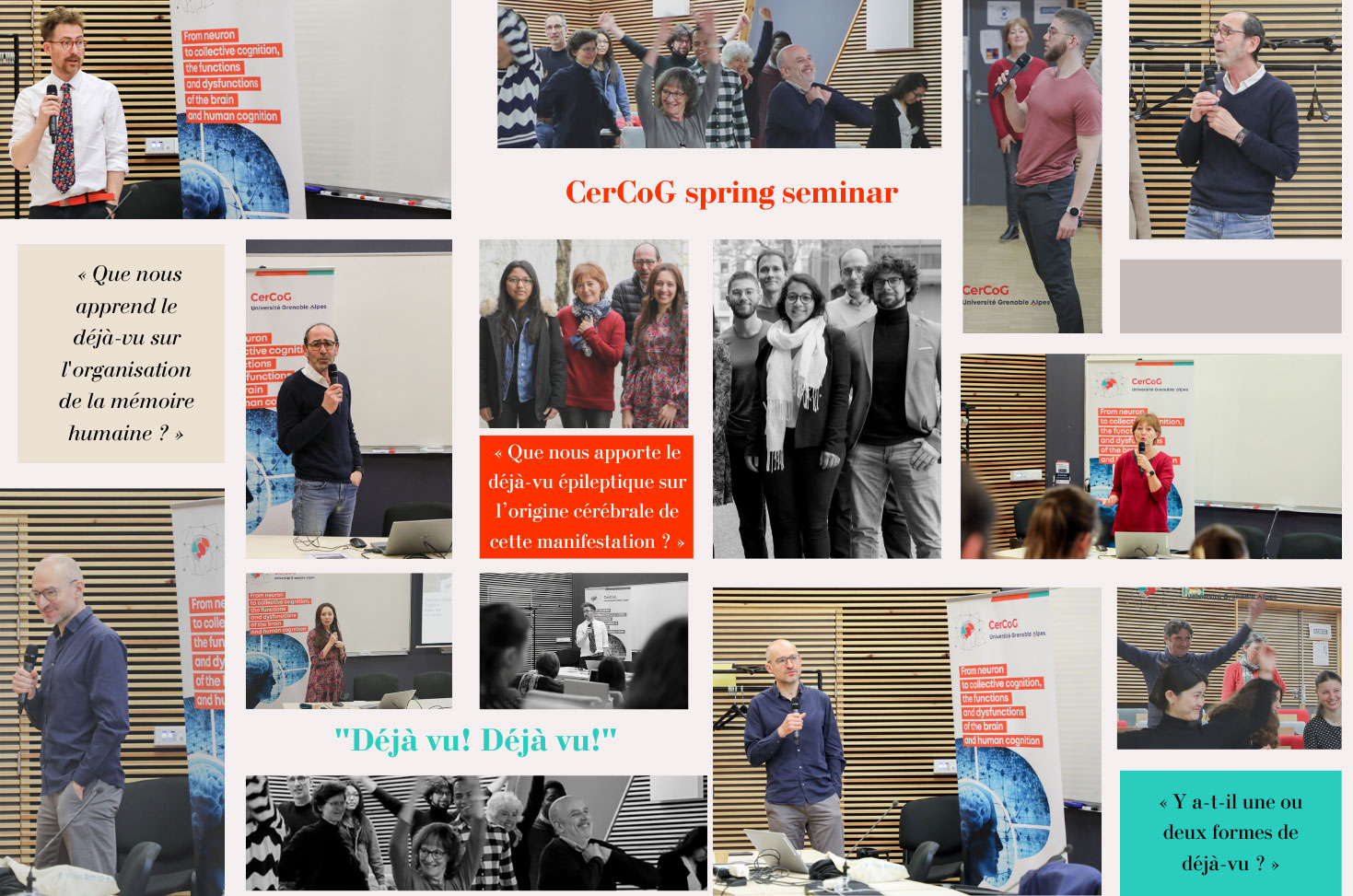
CerCoG's scientific events include seasonal seminars, scientific days and workshops.
Scientific junior council (COSJUN)
Composed of young researchers (PhD students, post-docs, young physicians) from various disciplines around the themes CerCog@UGA (Université Grenoble Alpes) thus recalling its three axes.
Scientific council (COS)
Composed of members representative of the scientific axes (4 members per scientific axis) and members of the Steering Committee led by the PI (principal investigator). It will regularly analyze and validate the content of scientific and methodological actions and the progress of the calls for projects.
TEAMS
CerCoG counts 595 members from 18 different labs structured in 3 axes.
Our teams belong to one of these axes:
Upcoming events
Call for projects
Bootstrapping
They are bootstrap projects to promote interdisciplinary interactions between teams within axes. The funding of this type of project serves to master internships and associated running costs. One call per year.
Collaborative
They are bootstrap projects to promote interdisciplinary interactions between teams, axes, and poles, providing a larger amount of funding for running costs and short-duration salaries. One call per year.
Structuring
They are federative projects to promote and develop CerCoG inter-axe collaborations between teams. Funding serves for running costs and salaries. One call for the duration of the program.
Platforms
Funding strengthens the CerCoG platform network and targets common methodological developments for our Brain and Cognition community.
Communication
Communication
A set of podcasts through which CerCoG plans, this season, to train its young researchers to communicate effectively.

595
CerCoG members
395
Permanent members
18
Labs
4
Poles